This technical note focuses on the development of a 3D model using GrowDex®-T in a Be-Doubleflow chip under active flow.
Introduction
Rapidly evolving fields of medicine and pharmaceutics require more robust, cost-effective, ethical, and complex systems to better understand the human body and to develop new treatments for debilitating diseases. It has been shown that commonly used animal models often fail to represent the human system [1]. In addition, the conventional in vitro setups, which do integrate human cells, face challenges in recreating the required complexity of the surroundings where the cells reside [2, 3].
Listed as one of the top 10 emerging technologies in 2016, organ-on-chips (OOCs) are in vitro platforms that promise to revolutionise the way cell models have been done in the past [4]. In general, OOCs are microfluidic cell cultivation devices that aim to recreate and sustain the micro and nanostructures (2D or 3D) of a chosen organ in their authentic environment. One of the benefits of OOC technology is the increased biological relevance from controlled cell culture conditions that extend to e.g., fluid flow, mechanical forces, and generation of gradients [5].
Aim of the study
The aim of this study was to establish a 3D environment using GrowDex®-T, an animal-free nanofibrillar cellulose hydrogel, in a commercial organ-on-chip device that could endure long-term active flow. We tested different GrowDex®-T concentrations in Be-Doubleflow chips and connected them to the flow from a peristaltic pump for 7 days. During these flow experiments, the overall performance and barrier integrity of GrowDex®-T were assessed.
Manufacturer recommendations state that the Be-Doubleflow must be incubated at 37°C, for a minimum of 24 hours before gel-loading to minimize the number of bubbles in the devices. Ideally, the whole experimental setup should be kept at a constant temperature because temperature changes have been found to increase the probability of bubble formation [6].
As an example, the most commonly used animal-derived hydrogels are prepared cold and gelate rapidly at higher temperatures [7]. Once the temperature-sensitive gel is loaded into warm chips, a significant temperature difference is introduced to the system which can induce the generation of bubbles. Furthermore, the loading of these gels can be challenging if the gelation occurs too fast and the designated microchannel is left only partially filled.
There are several benefits related to the use of GrowDex®-T in 3D cell culture. It can be loaded to chips at desired temperatures, thus bubble-inducing temperature variations can be greatly minimized. Quick and easy handling and preparation of GrowDex®-T dilutions, no need for cross-linking or gelation, and simple loading into the Be-Doubleflow system accelerates the workflow and efficient use of time for assay setup. Finally, the low autofluorescence and optical transparency of GrowDex®-T enables the use of both fluorescence and brightfield microscopy in the visualization of samples.
Materials
Organ-on-chip device
• Be-Doubleflow standard (Beonchip)
Biomaterial and fluorescent beads
• GrowDex®-T 10ml, stock: 1.0%, (Cat. No. 200 103 010, UPM Biomedicals)
• Fluoro-Max Green and Red Dry Fluorescent Particles, diameter: 15μm (Cat. No. 11895052, Thermo Scientific™)
Flow platform, connectors, tubing, and accessories
• Ismatec IP peristaltic pump, 12 channels
• tygon® Tubing 0.5mm ID x 1.5mm OD (PVC) (Cat. No. T4102, Qosina)
• HPLC tubing, Upchurch Scientific®, 1/16″ OD x 0.030″ ID x 100ft (Cat. No. 554-2980, VWR)
• Ferrules: Flangeless fittings for 1/16″ OD tubing, Upchurch Scientific® (Cat. No. UPCHP-200X, VWR)
• Nuts: Flangeless fittings for 1/16″ OD tubing, Upchurch Scientific® (Cat. No. UPCHXP-201X, VWR)
• Ismatec PharMed® BPT 0.19mm ID orange/red, 2-stop (Cat. No. WZ-95723-10, Cole-Parmer)
• Silicon tubing 1mm ID x 3mm OD (Cat. No. 228-0701, VWR)
Other reagents and laboratory accessories
• Working medium: DMEM, high glucose, no glutamine, Gibco™ (Cat. No. 11960044, ThermoFisher) with Dulbecco’s Phosphate Buffered Saline w/o Calcium w/o Magnesium (Cat. No. L0615, Biowest)
• Penicillin-Streptomycin (Cat. No. 11074440001, Roche)
• TipOne RPT 1250ul (Cat No. S1161-1820)
Microscope and image processing
• Zeiss Axio Observer.Z1
• Zeiss Zen (blue edition), version 3.3.89.0000
• Fiji-ImageJ-64bit, v1.52p
Methods
In this study, three GrowDex®-T concentrations (0.3%, 0.5% and 0.9%) were prepared and loaded into one channel of the Be-Doubleflow chip, then media was loaded into and pumped through the second channel, as active flow by peristaltic pump for 7 days. The layout and structure of the Be-Doubleflow are illustrated in Figure 1 and Figure 2. All preparations of GrowDex®-T dilutions and loadings into the Be-Doubleflow chips were done in a sterile environment following aseptic techniques. Green, fluorescent beads were suspended in GrowDex®-T to assess the barrier integrity of the samples. Qualitative analysis of GrowDex®-T was conducted using brightfield and fluorescence microscopy. GrowDex®-T barrier was considered intact if the fluorescent beads had not escaped with the flow or moved in the gel channel.
Images were taken from six time points: after loading GrowDex®-T and medium, after 24-hour incubation before flow initiation (static media); and 24 hours, 48 hours, 72 hours, and 7 days after the start of the flow. Furthermore, the first hour of flow was recorded real-time with the microscope. The experimental details of the study are summarized in Table 1. Beonchip technical data sheet, and technical support was provided by Beonchip for the handling of the chips [8, 9].
Table 1 Summary of flow test details using Be-Doubleflow devices
| Experimental conditions | Concentration of GrowDex®-T | Concentration of fluorescent beads | Flow platform | Flow rate |
| 1 | 0.3% | 1.5×10^6 beads/ml | Peristaltic pump | 12μl/min |
| 2 | 0.5% | 1.5×10^6 beads/ml | Peristaltic pump | 12μl/min |
| 3 | 0.9% | 1.5×10^6 beads/ml | Peristaltic pump | 12μl/min |
Preparing GrowDex®-T dilutions:
- GrowDex®-T dilutions (dilutions 0.3%, 0.5%, and 0.9% ) with fluorescent beads were prepared according to the GrowDex-T “Instructions for use” protocol [10] (the specific volumes used for the different GrowDex®-T concentrations are shown in Table 2). This can be repeated by following the protocol detailed below.
- Add DMEM (at room temperature – RT) into 1.5ml Eppendorf tube.
- Weigh GrowDex®-T (stock 1.0% at RT) into the Eppendorf tube on a scale (refer to table 2 for different dilutions).
- Mix the GrowDex®-T and DMEM thoroughly using a 1000μl pipette with low retention pipette tips until the solution appears homogeneous.
- Pipette 46.05μl of DMEM (at RT) into a separate 1.5ml Eppendorf tube.
- Mix in 3.95μl of green fluorescent beads (at RT) into 46.05μl of DMEM (final concentration: 1.5×10^6 beads/ml) thoroughly using a pipette.
- Add 50μl of bead suspension into all GrowDex®-T test concentrations (diluted 0.3%, 0.5%, and 0.9%).
- Mix the bead solution in GrowDex®-T thoroughly using a 1000μl pipette with low retention pipette tips so that the solution appears homogeneous.
Table 2 Dilution table for tested GrowDex®-T concentrations with fluorescent beads
| GrowDex®-T test concentration | Volume of DMEM | Volume of GrowDex®-T stock solution (1.0 %) | Volume of DMEM for bead suspension | Volume of beads -1 x DPBS solution (1.9 x 10^8 beads/ml) |
| 0.3 % | 300 µl | 150 µl | 46.05 µl | 3.95 µl |
| 0.5 % | 200 µl | 250 µl | 46.05 µl | 3.95 µl |
| 0.9 % | 0 µl | 500 µl | 46.05 µl | 3.95 µl |
Figure 1.
Graphical image illustrating the layout of the two microfluidic channels within the Be-Doubleflow chip.
Figure 2.
Top view of Be-Doubleflow chip:
A) Upper channel inlet and inlet reservoir;
B) upper channel medium overflow reservoir;
C) bottom channel inlet and inlet reservoir;
D) bottom channel medium overflow reservoir;
E) evaporation reservoir;
F) upper channel outlet and outlet reservoir,
G) bottom channel outlet and outlet reservoir. Modified from Be-Doubleflow technical characteristics data sheet.


Loading Be-Doubleflow with GrowDex®-T:
1. According to the manufacturer’s instructions, prior to addition of the hydrogel, empty Be-Doubleflow chips were incubated over the weekend at 37°C and 5% CO² in humidity chambers, i.e. large petri dishes with smaller petri dishes inside filled with H²O (Figure 3).
2. Add 400μl of 1 x DPBS into the evaporation reservoirs (Figure 2E).
3. Pipette 100μl of GrowDex®-T with fluorescent beads into the bottom channel from one side using a 1000μl pipette (Figure 2C).
4. Lastly, add 50μl of DMEM (37°C) into the bottom channel inlets to prevent drying (Figure 2C and G).
5. Incubate the GrowDex®-T filled chips at 37°C and 5% CO² in humidity chambers for 30 minutes.
6. To fill the top media channel, add 100μl of DMEM (37°C) into the upper channel using a 1000μl pipette (Figure 2A) until the media moves through the channel outlet (Figure 2G).
7. For the inlet and outlet reservoirs, add 300μl of DMEM (37°C), 100μl at a time, into the upper channel inlet reservoir (Figure 2A). The chip should be tilted between the medium additions to allow it to flow through to the outlet side of the chip (Figure 2F). Media may overflow into the medium overflow reservoirs.
8. After medium additions, Be-Doubleflow chips should be incubated at 37°C and 5% CO² in humidity chambers for 24 hours before starting the flow (Figure 3).
Figure 3.
Humidity chamber made with petri dishes for Be-Doubleflow chips.

Connecting Be-Doubleflow to the peristaltic pump flow:
1.Sterilize pump tubing by pumping through 70% ethanol and rinse by pumping through dH²O. Connector nuts and ferrules should be soaked in 70% ethanol on a rocker and rinsed by soaking them in dH²O on a rocker. In addition, nut interiors can be sterilized by injecting 70% ethanol and rinsed by injecting dH²O using a 10ml syringe.
2.Prime pump tubing with DMEM (37°C). The pump was set to minimum rate while fixing the chips, connectors and plugs to avoid trapping air into the flow system.
3.Screw the plugs onto the medium-filled bottom channel inlet and outlet reservoirs (Figure 2C and G).
4.Fill the top channel inlet and outlet reservoirs with DMEM (37°C).
5.Aspirate overflowing medium from the medium reservoirs (Figure 2B and D).
6.Connect primed pump tubing onto the top channel inlet reservoirs via connectors (Figure 2A).
7.Connect empty outlet tubing onto the top channel outlet reservoirs via connectors (Figure 2F).
8.Aspirate overflowing medium from the medium reservoirs (Figure 2B and D).
9.In these experiments, the pump was set to a working speed of 2.01rpm which is equal to 12μl/min (DMEM at 37°C).
10.For the first hour of flow, the pump and chips were assembled next to the fluorescent microscope and time lapse images were collected (one chip/condition).
11.After the first hour of flow, the chips setup (chips, tubing and media tubes) was moved into a humidified incubator (37°C, 5% CO²) to sustain the medium flow for 7 days (Figure 4). The chips were detached from the pump for imagining (Zeiss Axio Observer.Z1) at 24h, 48h, 72h and 7d of flow.
12. Subsequently taking the microscope images, the top channels, and inlet and outlet reservoirs were filled with DMEM (37°C) before re-connecting the tubing.
Figure 4.
Be-Doubleflow chips connected to a pump in a humidified incubator.

Results
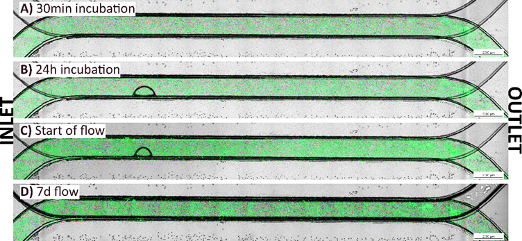
GrowDex®-T diluted to 0.3%, 0.5% and 0.9% was loaded successfully into the Be-Doubleflow chips (Figure 5, Figure 6, and Figure 7 respectively). The preparation of the GrowDex®-T concentrations was straightforward, and the beads were seen to be evenly distributed. All concentrations were fast and easy to pipet into the bottom channels using a 1000μl pipette (<1min/chip). GrowDex®-T filled the gel channels entirely, and no bubbles were observed after loading. (Figure 5A, Figure 6A, and Figure 7A) GrowDex®-T did not shrink or dry out after 30-minutes of incubation, or after the 24-hour incubation with static, non-flowing medium-filled top channel and reservoirs (Figure 5A-B, Figure 6A-B, and Figure 7A-B). Few bubbles were observed in GrowDex-T throughout the experiments (Figure 5, Figure 6, and Figure 7).
All GrowDex®-T concentrations withstood the 7 days of active flow (12μl/min) (Figure 5A, Figure 6A, and Figure 7A). No movement of beads were observed during the flow experiments. In addition, the beads did not leak from the bottom channels with the flow in the top channel. The beads were easy to visualize with a microscope while suspended in GrowDex®-T since the gels showed no turbidity or autofluorescence during imagining.
Figure 5.
0.3% GrowDex®-T loaded into a Be-Doubleflow chip over different time points with green fluorescent beads suspended in GrowDex®-T for visualization.
A) after medium addition;
B) 24h incubation;
C) immediately after flow initiation;
D) 7 days after flow initiation.
Flow rate: 12μl/min. Scale bar: 2mm.
Figure 6.
0.5% GrowDex®-T loaded into a Be-Doubleflow chip over different time points with green fluorescent beads suspended in GrowDex®-T for visualization.
A) after medium addition;
B) 24h incubation;
C) immediately after flow initiation;
D) 7 days after flow experiments.
Flow rate: 12μl/min. Scale bar: 2mm.
Figure 7.
0.9% GrowDex®-T loaded into a Be-Doubleflow chip over different time points with green fluorescent beads suspended in GrowDex®-T for visualization.
A) after medium addition;
B) 24h incubation;
C) immediately after flow initiation;
D) 7 days after flow experiments.
Flow rate: 12μl/min. Scale bar: 2mm.


Conclusions
Loading GrowDex®-T into the Be-Doubleflow chips is fast, easy, and simple. This was evidenced by the successful loading of all concentrations of GrowDex®-T with fluorescence beads, therefore it is expected that this could be repeated with live cells.
We found that using a 1000μl pipette is the best option for loading the GrowDex®-T. In addition, GrowDex®-T could endure incubations without signs of drying, shrinkage, or degradation in the conditions necessary for cell culture. Each GrowDex®-T sample showed very few bubbles during the entirety of the experiments. The flexibility of GrowDex®-T in terms of working temperature, likely has a positive impact on keeping the bubble formation under control.
However, some air pockets were introduced into some chips when loading the lower concentrations of GrowDex®-T (0.3% and 0.5%), whilst plugging the bottom channel inlets and outlets. Since lower concentrations of GrowDex®-T are not as stiff compared to the stock GrowDex®-T they deform easier with applied stress. Thus, we recommend screwing the plugs down gently when using lower GrowDex®-T concentrations to avoid introducing air pockets into the gels.
Regardless, GrowDex®-T gels remained unchanged during the seven days of active flow which supports their use in Be-Doubleflow setups with perfusion. Furthermore, since it is easy to successfully load a range of GrowDex®-T concentrations into the Be-Doubleflow chips, this enables the users to choose which concentration of GrowDex®-T will best suit their given application and cell type.
In conclusion, different GrowDex®-T concentrations were compatible with the Be-Doubleflow chips and are expected to be suitable in cell culture applications with perfusion.
AUTHORS
Elina Kalke¹, Kaisa Tornberg¹, Jonathan Sheard², Pasi Kallio¹
1 Tampere University, Faculty of Medicine and Health Technology, Tampere, Finland
² UPM Biomedicals, Helsinki, Finland
References
1.Van Norman, G., A. (2019). “Limitations of animal studies for predicting toxicity in clinical trials: Is it time to rethink our current approach?” JACC.Basic to Translational Science; JACC Basic Transl Sci, 4(7), 845-854.
2.Kapałczyńska, M., Kolenda, T., Przybyła, W., Zajączkowska, M., Teresiak, A., Filas, V., Lamperska, K. (2018). “2D and 3D cell cultures – a comparison of different types of cancer cell cultures.” Archives of Medical Science; Arch Med Sci, 14(4), 910-919.
3.Van der Meer, Anies Dirk, & van den Berg, A. (2012). Organs-on-chips: Breaking the in vitro impasse. Integrative Biology (Cambridge); Integr Biol (Camb), 4(5), 461-470.
4.Top 10 emerging technologies. (2016). Available online: https://www.weforum.org/agenda/2016/06/top-10-emerging-technologies-2016/, 16.6.2021
5.Wu, Q., Liu, J., Wang, X., Feng, L., Wu, J., Zhu, X., Gong, X. (2020). “Organ-on-a-chip: Recent breakthroughs and future prospects”. Biomedical Engineering Online; Biomed Eng Online, 19(1), 9-19.
6.Pereiro, I., Fomitcheva Khartchenko, A., Petrini, L., & Kaigala, G. V. (2019). ”Nip the bubble in the bud: A guide to avoid gas nucleation in microfluidics”. Lab on a Chip; Lab Chip, 19(14), 2296-2314.
7.Akther, F., Little, P., Li, Z., Nguyen, N., & Ta, H. T. (2020). “Hydrogels as artificial matrices for cell seeding in microfluidic devices”. RSC Advances, 1(71), 43682-4373.
8.BEOnChip. (2020). “Technical pdf”. Sourced from: https://beonchip.com/wp-content/uploads/2020/08/UserGuideBDF.pdf, 21.5.2021.
9.BEOnChip. (2020). “How to video”. Sourced from: Be-Doubleflow: Cell culture – YouTube, 21.05.2021.
10.UPM Biomedicals. (2020). GrowDex®-T “Instructions for use”.

